Medanta Hospital in Gurugram has achieved an important breakthrough in fetal therapy by successfully completing its first fetoscopic laser photocoagulation to treat Twin-to-Twin Transfusion Syndrome (TTTS). The complex intervention was performed under the leadership of Dr. Geetanjli Behl, Senior Consultant, Fetal Medicine, Obstetrics & Gynaecology, Medanta, Gurugram.
TTTS occurs only in monochorionic twin pregnancies, where both babies share a single placenta. Because the twins are connected through shared placental blood vessels, blood flows unevenly between them—one twin donates blood while the other receives excess circulation. If left untreated, the condition can lead to severe complications and even loss of one or both fetuses. Fetoscopic laser photocoagulation is globally recognized as the standard and most effective treatment.
A 30-year-old woman in her first pregnancy, carrying naturally conceived monochorionic diamniotic twins, arrived at Medanta at 20 weeks and 4 days of gestation after travelling from Doha. She had already been diagnosed with Stage II TTTS and sought definitive therapy through fetoscopic laser ablation of placental connecting vessels.
Doctors immediately performed a detailed antenatal ultrasound examination. The scan confirmed monochorionic diamniotic twins affected by Stage II TTTS. The fetuses showed a 17% difference in estimated weight. Additional findings included absence of the donor twin’s bladder, a marked difference in amniotic fluid levels, and abnormal Doppler readings — all consistent with Stage II disease.
The couple underwent comprehensive counselling. Doctors explained that without treatment, TTTS carries a 90–100% probability of losing one or both babies and that surviving infants may face significant neurological damage.
They were informed that fetoscopic laser photocoagulation improves the likelihood of survival of both twins to around 70% and guarantees at least one surviving baby in nearly 80% of cases. The procedure also lowers neurological complications to about 5%.
Possible risks were discussed as well, including premature rupture of membranes, preterm delivery, and about a 12% chance of persistent TTTS or progression to Twin Anaemia–Polycythaemia Sequence (TAPS). After understanding the risks and benefits, the couple consented to proceed.
The team carried out fetoscopic laser ablation using the Solomon technique, identifying and sealing roughly six placental vascular connections.
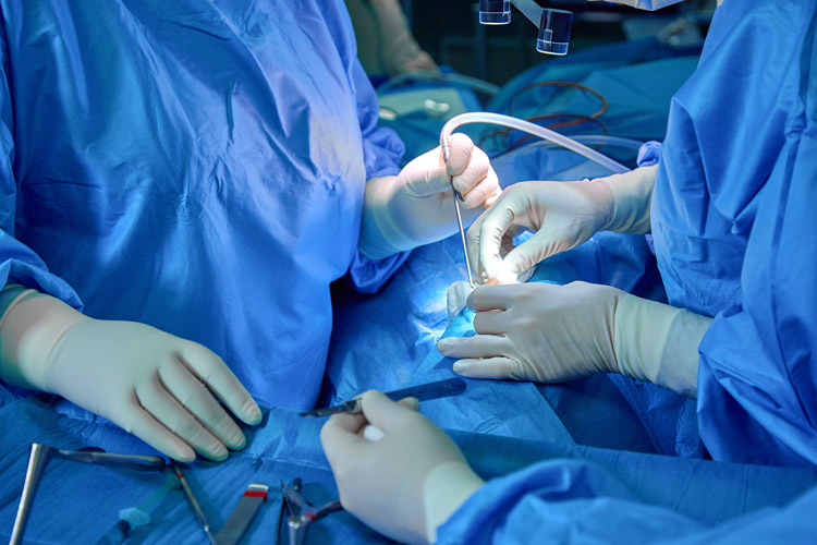
This minimally invasive surgery was performed under local anaesthesia. Guided continuously by ultrasound, surgeons selected an appropriate entry point to visualise the vascular equator of the placenta. A small incision was made in the mother’s abdomen, and a 10-French cannula was inserted into the uterus using the Seldinger method. A 3.3-mm curved fetoscope was then advanced to inspect the placental vessels. Using a 0.6-mm diode laser fibre, the abnormal vessel connections were coagulated.
To further reduce recurrence and minimise the risk of TAPS, the surgeons joined the coagulated points along the placental vascular equator in a continuous line — the hallmark of the Solomon technique.
After the procedure, around 1,300 ml of excess amniotic fluid was drained from the recipient twin’s sac. Both babies had stable heartbeats at the conclusion of surgery.
Post-procedure, the mother remained stable with no vaginal fluid leakage or abdominal pain. Ultrasound scans soon showed improvement. By the second postoperative day, the donor twin’s bladder became visible and normal amniotic fluid began accumulating.
The patient was discharged on the third day in good condition without contractions, pain, or leakage. Ultrasound evaluation at discharge confirmed adequate fluid around both fetuses and reassuring blood-flow patterns. Doctors advised weekly ultrasound checks for two weeks, followed by a detailed neurosonogram after four weeks.
The pregnancy continued safely until 34 weeks. She delivered two healthy boys weighing 2.2 kg and 2.0 kg. The smaller baby required brief NICU care, and both infants showed positive outcomes overall.
(The above image is for illustrative purposes only)