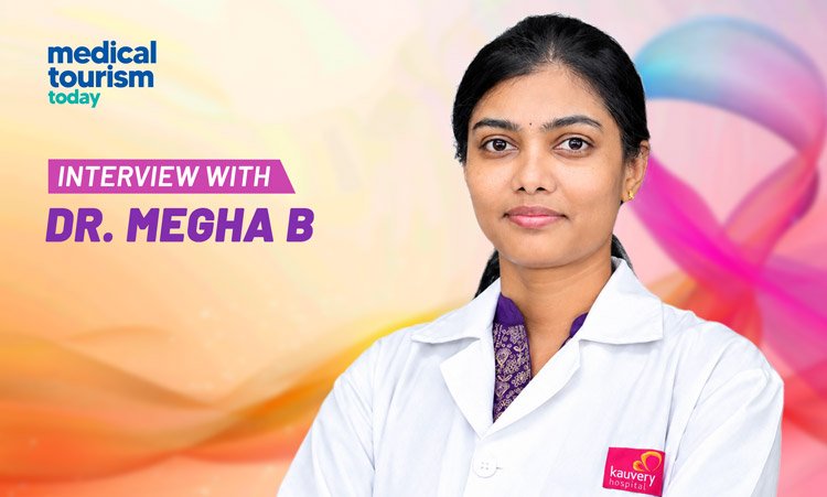
In an interview with Medical Tourism Today, Dr. Megha B, Surgical Oncologist and Consultant Breast Surgeon at Kauvery Hospital, Tennur, Trichy, shares expert insights into breast cancer awareness, early detection, individual risk assessment and advances in breast-conserving surgery. She also discusses the emerging role of artificial intelligence and robotic technologies in breast cancer care, emphasising the importance of timely diagnosis, personalised treatment and long-term breast health.
Breast-related symptoms such as lumps, pain, nipple discharge and changes in breast appearance are common, but they can also cause considerable anxiety. Which symptoms or changes should women never ignore, and when should they consult a breast specialist?
Most breast lumps and changes are benign. However, it is important to remember that a lump cannot be reliably classified as benign or malignant by touch alone. Although breast pain alone is rarely a sign of cancer, breast cancer does not always cause pain.
Women should consult a breast specialist, particularly if a change persists or does not settle after the menstrual cycle. They should also seek medical advice if they notice a new or persistent lump in the breast or armpit, especially if it is hard, increasing in size or associated with other changes. Other warning signs include an unexplained change in breast size or shape, new nipple inversion or a change in nipple direction, and spontaneous nipple discharge, particularly if it is clear or blood-stained and comes from one breast. Persistent crusting, scaling or other changes around the nipple should also be evaluated.
Skin changes such as dimpling, puckering, thickening, persistent redness or swelling, and an orange-peel appearance also warrant medical evaluation.
The key message for women is to recognise what is normal for their breasts and seek medical attention whenever they notice a new, unusual or persistent change.
Many women immediately associate a breast lump with cancer, while others may delay evaluation because they assume it is harmless. How do you assess a breast lump, and what role do clinical examination, imaging and image-guided biopsy play in reaching an accurate diagnosis?
Assessing a breast lump requires a combination of clinical judgement, appropriate imaging and, when indicated, a biopsy. This approach is known as triple assessment. During the clinical assessment, the doctor takes the patient’s medical history and examines both breasts, the nipples, areolae, underarm areas and relevant lymph node regions. The doctor assesses the lump’s location, size, consistency and mobility.
The choice of breast imaging depends on the woman’s age, symptoms, breast density and clinical findings. An ultrasound and a mammogram are commonly used to evaluate a lump, while an MRI is reserved for selected situations. If the examination or imaging identifies a suspicious abnormality, tissue sampling may be recommended. This may involve fine-needle aspiration cytology (FNAC) or a core needle biopsy, also known as a true-cut biopsy.
Access to screening, diagnostic and treatment facilities is not the only consideration. Many women find it difficult or uncomfortable to discuss breast symptoms, particularly with male doctors. This is precisely why we have established a clinic at Maa Kauvery Breast Clinic, Trichy, that is staffed entirely by women experts. Patients can discuss all aspects of breast cancer in a supportive environment.
Early detection can significantly influence breast cancer treatment and outcomes. What should women know about breast screening, breast self-awareness and individual risk assessment, and how should screening approaches vary according to age, family history and personal risk factors?
Early detection plays an important role in the timely treatment of breast cancer. However, women should understand that breast self-awareness, screening and diagnostic evaluation are three distinct aspects of breast health.
Breast self-awareness means becoming familiar with how one’s breasts normally look and feel, including their usual size, shape, texture and nipple appearance. This helps women recognise new or persistent changes without repeatedly examining their breasts or following a complicated self-examination technique.
Screening is intended for women who do not have symptoms. It aims to detect breast cancer before it becomes clinically apparent. Mammography is an established screening tool, but the appropriate starting age and screening interval depend on the woman’s age, individual risk, breast density and other factors. Women should therefore discuss an appropriate screening plan with their breast specialist rather than follow a one-size-fits-all schedule.
Diagnostic evaluation, on the other hand, is undertaken when a woman has a breast symptom or when a screening test reveals an abnormality requiring further investigation. For example, a woman who notices a new lump, nipple discharge, changes in breast skin or a newly inverted nipple should consult a doctor, even if she is younger than the recommended screening age or has recently had a normal mammogram.
Depending on the clinical findings, the evaluation may include a clinical breast examination, diagnostic mammography, ultrasound or other imaging tests. If a suspicious abnormality is identified, a biopsy may be recommended to determine whether it is cancerous. Unlike screening, diagnostic evaluation is intended to investigate a specific symptom or finding and establish a diagnosis.
Family history is a major concern for many women. How much can a family history of breast cancer influence an individual’s risk, and when should women consider genetic counselling or a more detailed assessment of their breast cancer risk?
Family history is an important factor in assessing a woman’s risk of breast cancer. However, the degree of risk varies according to the number of affected relatives, their relationship to the woman, their ages at diagnosis and whether cancers such as breast and ovarian cancer occur on the same side of the family. Both the mother’s and the father’s sides of the family are equally relevant because inherited cancer-predisposition variants can be passed down through either parent.
Women should discuss their family history with a doctor, particularly if their mother, sister or daughter has had breast cancer; if relatives were diagnosed at a young age; if multiple family members have had breast or ovarian cancer; if breast cancer has affected both breasts; or if there is a history of male breast cancer or a known genetic mutation in the family. These patterns may indicate an increased likelihood of hereditary cancer susceptibility and the need for a more detailed assessment.
Genetic counselling may be considered when a woman’s personal or family history suggests an inherited risk. BRCA1 and BRCA2 are among the best-known genes associated with hereditary breast cancer, although other genes may also be involved. Women identified as having an increased risk may need an individualised screening and surveillance plan.
The most important message is that family history helps identify women who may need additional assessment, but having a family history does not mean that a woman will definitely develop the disease. Similarly, the absence of a family history does not mean that a woman is free from risk. In fact, more than 80% of women diagnosed with breast cancer have no known family history. A woman’s individual risk depends on a combination of factors, including age, reproductive and hormonal history, lifestyle, previous breast conditions and genetic susceptibility.
You specialise in breast-conserving and oncoplastic breast surgery. How have these approaches transformed breast cancer surgery, and how do you balance effective cancer removal with preservation of breast shape, appearance and quality of life?
A breast cancer diagnosis does not necessarily mean that the entire breast must be removed. Breast-conserving and oncoplastic surgery have changed the way we approach breast cancer treatment by shifting the focus from simply removing the tumour to treating the cancer effectively while also considering the woman’s long-term physical and emotional well-being.
The balance between effective cancer removal and preservation of appearance depends on several factors, including the tumour’s size, stage and biology, the characteristics of the breast and the patient’s preferences.
In breast-conserving surgery, the priority is always the safe removal of the cancer while preserving the breast and achieving a good cosmetic outcome wherever possible. Surgical planning must therefore be individualised rather than follow a one-size-fits-all approach.
Oncoplastic surgery goes beyond appearance alone. It allows surgeons to consider both cancer control and the physical outcome of surgery, with the aim of preserving breast shape, symmetry and the patient’s quality of life.
The goal is to achieve the best possible balance between oncological safety, good cosmetic outcomes and the patient’s overall well-being.
With AI and robotic technologies increasingly being explored in cancer care, how do you see them contributing to the future of breast cancer treatment? Could these technologies improve surgical precision, treatment planning, patient outcomes and recovery, and what are their current limitations in breast surgery?
Artificial intelligence and robotic technologies have considerable potential to improve breast cancer care, from diagnosis and treatment planning to surgery and recovery. However, they serve different purposes: AI analyses information to support clinical decisions, while robotic systems assist surgeons in performing procedures. Neither replaces the expertise and judgement of the treating team.
AI is already being used in breast imaging and is being explored in digital pathology, treatment planning and outcome prediction. It can help identify suspicious areas on mammograms, analyse tumour characteristics and integrate imaging, pathology and clinical information to support more personalised treatment decisions. In radiotherapy, AI can help clinicians plan treatment more efficiently and consistently. Its role in surgical planning, real-time guidance and the assessment of surgical margins is promising but still requires further validation.
Robotic surgery offers a different set of potential benefits. Three-dimensional visualisation, articulated instruments and improved access to difficult-to-reach areas may help surgeons perform selected procedures through smaller incisions. In breast cancer care, robotic techniques are being explored particularly in nipple-sparing mastectomy and breast reconstruction, where incision placement and cosmetic outcomes are important. For appropriately selected patients, these approaches may offer aesthetic advantages, although the extent of improvement in recovery and complication rates varies.
Even after successful treatment, many breast cancer survivors have questions about recurrence, follow-up, lifestyle and returning to normal life. What does comprehensive post-treatment surveillance involve, and what should survivors keep in mind for their long-term breast health?
Completing treatment is not the end of care; it is the beginning of a new phase of health management. Follow-up should be individualised, symptoms should not be ignored, and survivors should feel empowered to discuss physical, emotional and lifestyle concerns with their healthcare team.
Women with a personal history of breast cancer may require an individualised surveillance plan, including appropriate breast imaging and regular medical follow-up. Long-term follow-up is important for monitoring breast health, identifying concerning changes and providing ongoing support. The goal is not merely to detect recurrence but also to help women live healthier, fuller lives after breast cancer.
Survivors should remain familiar with how their breasts normally look and feel and seek medical advice if they notice a new lump, changes in breast size or shape, nipple discharge, nipple inversion, skin dimpling, or persistent redness or swelling.
Lifestyle also remains important. Regular physical activity, maintaining a healthy weight, limiting or avoiding alcohol, avoiding tobacco and attending recommended follow-up appointments support overall health.
The key message is that breast health requires continued attention even after successful treatment. An individualised follow-up plan, awareness of new symptoms and timely medical evaluation can help survivors manage their long-term health with greater confidence.
There are numerous myths and misconceptions surrounding breast cancer, including those related to lifestyle, breastfeeding, breast injuries, bras and other everyday factors. Which misconceptions would you most like to correct, and what is the most important message you would like every woman to remember about taking charge of her breast health?
One of the most important misconceptions I would like to correct is the belief that a woman cannot develop breast cancer if there is no family history of the disease. This is not true. Many women diagnosed with breast cancer have no known family history, and the absence of a family history does not mean there is no risk.
Another misconception is that every breast lump is cancerous. Many breast lumps, particularly in younger women, are benign conditions such as fibroadenomas and cysts. However, a new or persistent lump should always be evaluated by a qualified healthcare professional rather than assumed to be harmless.
I would also like women to understand that breast cancer does not always cause pain. A painless lump or an unexplained change in breast shape, nipple appearance or skin texture should not be ignored. Similarly, having a risk factor does not mean that a woman will definitely develop cancer, and not having a known risk factor does not guarantee protection.
The most important message I would like every woman to remember is that breast awareness is not about fear; it is about knowing your body and knowing when to act. Become familiar with how your breasts normally look and feel, recognise new or persistent changes, seek timely medical advice and discuss an appropriate screening plan based on your age and individual risk. Early recognition, accurate diagnosis and appropriate treatment matter.
Dr. Megha B is a Surgical Oncologist and Consultant Breast Surgeon at Kauvery Hospital, Trichy. She holds an MCh in Surgical Oncology from Kidwai Memorial Institute of Oncology, Bengaluru, an MS in General Surgery from Mysore Medical College & Research Institute, and an MBBS from Basaveshwara Medical College & Research Centre.
She has worked as a Senior Resident at Shimoga Institute of Medical Sciences and as an Assistant Professor at ESIC Medical College & Hospital, Rajajinagar, Bengaluru. She is also an Intuitive-certified robotic surgery first assistant, with experience in robotic-assisted surgery.
Dr. Megha has published research in diabetic wound care and facial reconstruction. She follows an evidence-based, research-oriented approach to patient care.