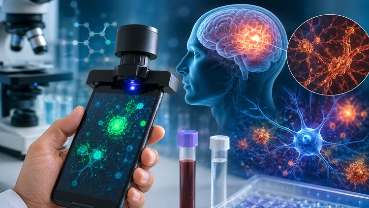
A research team at the Jawaharlal Nehru Centre for Advanced Scientific Research (JNCASR) has developed a portable fluorescence-based system that uses a smartphone to detect amyloid-β (Aβ), a biomarker strongly associated with Alzheimer’s disease.
The technology combines a newly developed fluorescent molecule, TZ-48, with a smartphone-enabled platform called ADxFluor. The approach is designed to measure changes in fluorescence produced when TZ-48 interacts with Aβ.
A New Approach to Biomarker Detection
Alzheimer’s disease gradually affects memory, thinking and behaviour. One of its major biological features is the build-up of amyloid-β proteins, which form fibrils and plaques in the brain. Detecting such biomarkers can help researchers assess disease-related changes.
Conventional techniques such as PET scans and MRI provide valuable clinical information but depend on expensive equipment and specialised infrastructure. Tests involving blood and cerebrospinal fluid can offer alternative ways to study Aβ and tau, but these methods may also require sophisticated laboratory facilities.
The JNCASR researchers therefore focused on developing a detection method that could be more portable, practical and scalable.
Fluorescence Probe Targets Aβ
TZ-48 was engineered to recognise Aβ fibrils and produce measurable fluorescence changes. The probe generates both increased fluorescence intensity and characteristic fluorescence lifetime signals when it interacts with the target.
Fluorescence lifetime measurements can have an advantage over conventional intensity-based analysis because they are comparatively less affected by probe concentration and photobleaching.
The findings, published in ACS Chemical Neuroscience, showed that TZ-48 could cross the blood-brain barrier and bind to Aβ plaques in transgenic mouse models of Alzheimer’s disease.
Researchers also tested the probe using confocal laser scanning microscopy and fluorescence lifetime imaging microscopy (FLIM). These techniques enabled quantitative assessment of Aβ accumulation at different stages of disease progression.
Detection in Blood and Cerebrospinal Fluid
The study went beyond brain imaging. TZ-48 was also able to detect Aβ in cerebrospinal fluid and blood serum. In experiments, the technology helped distinguish Alzheimer’s disease model mice from healthy animals.
To bring the detection process into a more portable format, Krithi K. Bhagavath, Madhu Ramesh, Yogendra Kumar, Sabyasachi Mandal, Hiriyakkanavar Ila and Thimmaiah Govindaraju developed ADxFluor.
The smartphone-integrated platform analyses the fluorescence generated by TZ-48 and uses it to quantify Aβ levels in serum. This combination of a molecular probe, fluorescence analysis and smartphone technology could open the way for simpler biomarker testing.
Potential for Portable Diagnostics
According to the researchers, the technology brings together three capabilities: imaging Aβ in brain tissue, detecting the biomarker in biological fluids and analysing fluorescence through a smartphone.
The platform could eventually serve as a foundation for portable diagnostic technologies targeting Alzheimer’s disease and potentially other neurodegenerative disorders.
The research is, however, still in the preclinical stage. The findings have been demonstrated in laboratory and animal studies, and human clinical trials will be needed to determine the system’s accuracy, reliability and suitability for real-world diagnostic use.
The JNCASR research demonstrates how smartphone technology and advanced fluorescent probes could be combined to make biomarker analysis more portable. While substantial clinical validation remains ahead, the approach represents a promising step towards developing simpler and more accessible tools for Alzheimer’s disease detection.