By Dr. Digish C. Shah, Senior Consultant – Nuclear Medicine, KD Hospital.
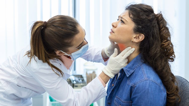
A patient comes into the clinic with a lump in the neck. It has been there for some weeks, growing slowly, not causing any fever, and not really hurting. A troubling thought arises: an ultrasound reveals a lymph node, and a biopsy confirms cancer. The harder question follows: where did it originate, and has it spread elsewhere?
Cancer can sometimes spread in ways that are difficult to predict. A tumour that appears small and limited to one area may have already spread to nearby lymph nodes in the neck. In some cases, the primary tumour may remain hidden and is discovered only after cancer is found elsewhere in the body.
This is where PET-CT can play an important role. Rather than looking only at the size or appearance of a tumour, PET-CT also shows how tissues are behaving, including areas with increased metabolic activity that may suggest cancer. And because it can assess the body in a single scan, PET-CT can help doctors look for disease beyond the site where it was first detected.
At KD Cancer Centre, this tool is one of the tools used in our head and neck cancer program. It does not replace the doctor’s thinking or biopsies. It often finds things that other methods do not.
What Is PET CT?
PET CT is really two scans working together. The PET part uses a safe amount of a radioactive substance. Usually FDG, a type of glucose. That is put into the blood. Cancer cells often take in glucose faster than normal cells. So they take in more of the substance. Show up as bright spots on the scan. The CT part, done at the time, gives a detailed picture of the body showing exactly where those bright spots are.
Neither scan alone tells the story. A CT scan can show a mass. Not always say if it is cancer or just scar tissue. A PET scan can show activity but not always know where it is. When they are put together, PET CT does both. It shows where something is and what it might be doing.
Why This is So Important in Head and Neck Cancer
Finding a Main Tumour
When cancer is found in a neck lymph node, but the source can’t be found through checking or looking inside, doctors call this a “cancer of unknown primary.” It is more common than people think. It used to mean patients had to have biopsies in the mouth and throat. PET-CT changed that. It often detects the main tumour because it shows areas of activity throughout the body. This lets biopsy target the spot, not guess.
Showing How Lymph Nodes Are Affected
Head and neck cancers often move to lymph nodes early. The problem is that a node can have cancer but still look normal on a CT or MRI scan. PET-CT can detect some of these cases by identifying increased metabolic activity associated with cancer, even before structural changes become clearly visible. This information can help doctors determine the most appropriate treatment approach, which may include neck surgery, radiation therapy, or a combination of both.
Finding Cancer That Has Spread
PET-CT looks at the body, not just the neck. This helps find cancer in the lungs, liver or bones. This is very important for treatment. A patient with cancer in the head and neck might get surgery or radiation. Someone with cancer might not. Knowing this before starting treatment helps avoid procedures and gets the right care.
Finding a Second Unrelated Cancer
People with a history of using tobacco or alcohol. Risk factors for head and neck cancer. Have a higher chance of getting a second separate cancer, often in the lung or oesophagus. Because PET- CT scans examine the body, they often detect second cancers when they are easiest to treat.
Helping Radiation Be Precise
The head and neck area has parts that patients need to keep working. Salivary glands, the spinal cord, and muscles used for swallowing. Radiation doctors use PET-CT to find the edges of the tumour. This means the right amount of radiation is used to control the cancer while keeping the parts safe.
Telling Scar Tissue from a Return of Cancer
Surgery and radiation change the neck. Scars and thickening changes that look like cancer on a CT or MRI. PET-CT helps by showing if the area is active, which is the way to tell if it is real cancer or just the result of treatment.
What to Expect During the Scan?
The process is simple. Patients are asked not to eat for an hour. They also avoid exercise the day before, as muscle activity can change how the substance spreads. After the FDG tracer is injected, there is a waiting period of about 45–60 minutes to allow it to distribute through the body. The scan typically lasts between 20 and 30 minutes. During the scan, patients should lie still and remain comfortable. Most people go back to their day after. The substance leaves the body naturally in 24 hours.
Head and neck cancer often hides its size at first. What looks like a problem may involve hidden spread, a hidden main tumour, or cancer that has gone elsewhere. PET-CT helps doctors see beyond the tumour and understand the whole disease. This information changes many of the choices in a patient’s care.
At KD Cancer Centre, we see PET-CT not as a standalone answer, but as an important part of a comprehensive approach to treating head and neck cancer. By combining PET-CT findings with other clinical and diagnostic information, our doctors can better understand the extent and activity of the disease and plan treatment accordingly.

Dr. Digish C. Shah is a Senior Consultant in Nuclear Medicine at KD Hospital, with more than 10 years of experience in molecular imaging and nuclear therapy, with a special focus on oncology. He holds an MBBS and DNB in Nuclear Medicine and has expertise in diagnostic molecular imaging, oncological and non-oncological PET-CT, Gamma Camera/SPECT imaging, radionuclide therapies and theranostic procedures.
Dr. Shah has published research articles in national and international journals and has served as faculty at several conferences in Nuclear Medicine. He is also a lifetime member of professional organisations, including SNMI, ANMPI, NAMS and IASO.